


The time between exposure to the virus and the development of symptoms of the disease is usually 2 to 21 days Estimates based on mathematical models predict that around 5% of cases may take greater than 21 days to develop
Symptoms usually begin with a sudden influenza-like stage characterized by feeling tired, fever, pain in the muscles and joints, headache, and sore throatThe fever is usually greater than 38.3 °C (100.9 °F).This is often followed by: vomiting, diarrhea and abdominal pain Shortness of breath and chest pain may occur next along with swelling, headaches and confusion.[12] In about half of cases the skin may develop a maculopapular rash (a flat red area covered with small bumps).[
 In some cases, internal and external bleeding may occur.[1] This typically begins five to seven days after first symptoms.[14] All people show some decreased blood clotting.[13] Bleeding from mucous membranes or from sites of needle punctures is reported in 40–50% of cases.[15] This may result in the vomiting of blood, coughing up of blood, or blood in stool.[16] Bleeding into the skin may create petechiae, purpura, ecchymoses, or hematomas (especially around needle injection sites).[17] There may also be bleeding into the whites of the eyes. Heavy bleeding is uncommon and if it occurs is usually within the gastrointestinal tract
Recovery may begin between 7 and 14 days after the start of symptoms.[12] Death, if it occurs, is typically 6 to 16 days from the start of symptoms and is often due to low blood pressure from fluid loss.[2] In general, the development of bleeding often indicates a worse outcome and this blood loss can result in death.[11] People are often in a coma near the end of life.[12] Those who survive often have ongoing muscle and joint pain, liver inflammation, and decreased hearing among other difficulties
Cause
Main articles: Ebolavirus (taxonomic group) and Ebola virus (specific virus)
Ebola virus disease in humans is caused by four of five viruses in the genus Ebolavirus. The four are Bundibugyo virus (BDBV), Sudan virus (SUDV), Taï Forest virus (TAFV), and one called, simply, Ebola virus (EBOV, formerly Zaire Ebola virus).[19] Ebola virus is the only member of the Zaire ebolavirus species and the most dangerous of the known EVD-causing viruses, as well as being responsible for the largest number of outbreaks.[20] The fifth virus, Reston virus (RESTV), is not thought to cause disease in humans, but has caused disease in other primates.[21][22] These five viruses are closely related to marburgviruses.[19]
Transmission
Life cycles of the Ebolavirus
The spread of Ebola between people occurs only by direct contact with the blood or body fluids of a person after symptoms have developed.[1][3] Body fluids that may contain ebolaviruses include saliva, mucus, vomit, feces, sweat, tears, breast milk, urine, and semen.[23] Entry points include the nose, mouth, eyes, or open wounds, cuts and abrasions.[23] Contact with objects contaminated by the virus, particularly needles and syringes may also transmit the infection.[24] The virus is able to survive on objects for a few hours in a dried state and can survive for a few days within body fluids.[23] Ebola virus may be able to persist in the semen of survivors for up to seven weeks after recovery, which could give rise to infections via sexual intercourse.[1] Otherwise, people who have recovered are not infectious.[24] The potential for widespread infections in countries with medical systems capable of observing correct medical isolation procedures is considered low.[25] Usually when someone has symptoms, they are sufficiently unwell that they are unable to travel without assistance.[26]
Handling infected dead bodies is a risk, including embalming.[25] Because dead bodies are still infectious, traditional burial rituals may spread the disease. Nearly two thirds of the cases of Ebola infections in Guinea during the 2014 outbreak are believed to have been contracted via unprotected (or unsuitably protected) contact with infected corpses during certain Guinean burial rituals.[27][28]
Healthcare workers treating those who are infected are at greatest risk of disease.[24] This occurs when they do not wear appropriate protective clothing such as masks, gowns, gloves and eye protection.[24] This is particularly common in parts of Africa where the health systems function poorly and where the disease mostly occurs.[29] Hospital-acquired transmission has also occurred in African countries due to the reuse of needles.[30][31] Some healthcare centers caring for people with the disease do not have running water.[32] In the United States, spread has occurred due to inadequate isolation.[33]
Airborne transmission has not been documented during EVD outbreaks.[3] Transmission among rhesus monkeys via breathable 0.8–1.2 μm aerosolized droplets has been demonstrated in the laboratory.[34] That airborne transmission does not appear to occur in humans may be due to there not being high enough levels of the virus in the lungs.[35] Spread by water or food other than bushmeat has also not been observed,[24] nor has spread by mosquitos or other insects.[24]
Initial case
Bushmeat being prepared for cooking in Ghana, 2013. Human consumption of equatorial animals in Africa in the form of bushmeat has been linked to the transmission of diseases to people, including Ebola.[36]
While it is not entirely clear how Ebola initially spreads from animals to human, it is believed to involve direct contact with an infected wild animal or fruit bat.[24] Wild animals other than bats capable of being infected include: a number of monkey, chimpanzees, gorillas, baboons and duikers.[37] In Africa wild animals, including fruit bats are hunted to eat, being known as bushmeat.[38][39]
Animals may become infected when they eat fruit already partially eaten by bats carrying the virus.[40] Fruit production, animal behavior, and other factors may trigger outbreaks among animal populations.[40]
It does appear that both domestic dog and pigs can also be infected with ebola viruses.[41] Dogs when they carry the virus do not appear to develop symptoms, while pigs appear to be able to transmit the virus to at least some primates.[41]
Reservoir
The natural reservoir for Ebola has yet to be confirmed; however, bats are considered to be the most likely candidate.[42] Three types of fruit bats (Hypsignathus monstrosus, Epomops franqueti, and Myonycteris torquata) have been found to possibly carry the virus without getting sick.[40][43] Whether or not other animals are involved in its spread is not known as of 2013.[41] Plants, arthropods, and birds have also been considered as possible reservoirs as well.[1]
Bats were known to reside in the cotton factory in which the first cases of the 1976 and 1979 outbreaks were observed, and they have also been implicated in Marburg virus infections in 1975 and 1980.[44] Of 24 plant species and 19 vertebrate species experimentally inoculated with EBOV, only bats became infected.[45] The bats displayed no clinical signs and is evidence that these bats are a reservoir species of the virus. In a 2002–2003 survey of 1,030 animals including 679 bats from Gabon and the Republic of the Congo, 13 fruit bats were found to contain EBOV RNA fragments.[46] Antibodies against Zaire and Reston viruses have been found in fruit bats in Bangladesh, thus identifying potential virus hosts and signs of the filoviruses in Asia.[47]
Between 1976 and 1998, in 30,000 mammals, birds, reptiles, amphibians and arthropods sampled from outbreak regions, no Ebola virus was detected apart from some genetic traces found in six rodents (Mus setulosus and Praomys) and one shrew (Sylvisorex ollula) collected from the Central African Republic.[44][48] Further efforts; however, have not confirmed rodents as a reservoir.[49] Traces of EBOV were detected in the carcasses of gorillas and chimpanzees during outbreaks in 2001 and 2003, which later became the source of human infections. However, the high lethality from infection in these species makes them unlikely as a natural reservoir.[44]
Virology
Main articles: Ebolavirus (taxonomic group) and Ebola virus (specific virus)
Electron micrograph of an Ebola virus virion
Ebolaviruses contain single-strand, non-infectious RNA genomes.[50] Ebolavirus genomes are approximately 19 kilobase pairs long and contain seven genes in the order 3'-UTR-NP-VP35-VP40-GP-VP30-VP24-L-5'-UTR.[51] The genomes of the five different ebolaviruses (BDBV, EBOV, RESTV, SUDV, and TAFV) differ in sequence and the number and location of gene overlaps. Like all filoviruses, ebolavirions are filamentous particles that may appear in the shape of a shepherd's crook or in the shape of a "U" or a "6", and they may be coiled, toroid, or branched.[51] In general, ebolavirions are 80 nanometers (nm) in width and may be as long as 14,000 nm.[52] In general, the median particle length of ebolaviruses ranges from 974 to 1,086 nm (in contrast to marburgvirions, whose median particle length was measured at 795–828 nm), but particles as long as 14,000 nm have been detected in tissue culture.[53]
Their life cycle begins with virion attachment to specific cell-surface receptors, followed by fusion of the virion envelope with cellular membranes and the concomitant release of the virus nucleocapsid into the cytosol. Ebolavirus' structural glycoprotein (known as GP1,2) is responsible for the virus' ability to bind to and infect targeted cells.[54] The viral RNA polymerase, encoded by the L gene, partially uncoats the nucleocapsid and transcribes the genes into positive-strand mRNAs, which are then translated into structural and nonstructural proteins. The most abundant protein produced is the nucleoprotein, whose concentration in the cell determines when L switches from gene transcription to genome replication. Replication results in full-length, positive-strand antigenomes that are, in turn, transcribed into negative-strand virus progeny genome copy. Newly synthesized structural proteins and genomes self-assemble and accumulate near the inside of the cell membrane. Virions bud off from the cell, gaining their envelopes from the cellular membrane they bud from. The mature progeny particles then infect other cells to repeat the cycle. The Ebola virus genetics are difficult to study due to its virulent nature.[55]
Pathophysiology
Pathogenesis schematic
Cells lining the inside of blood vessels (endothelial cells), macrophages, monocytes, and liver cells are the main targets of infection. Macrophages are the first cells to be infected with the virus and this infection results in cellular death.[52] Endothelial cells can be infected within three days after exposure to the virus.[52] After infection, a secreted glycoprotein, known as small soluble glycoprotein (sGP) or as the Ebola virus glycoprotein (GP), is synthesized. Ebolavirus replication overwhelms protein synthesis of infected cells and host immune defenses. The GP forms a trimeric complex, which binds the virus to the endothelial cells. The sGP forms a dimeric protein that interferes with the signaling of neutrophils, a type of white blood cell, which allows the virus to evade the immune system by inhibiting early steps of neutrophil activation. These white blood cells also serve as carriers to transport the virus throughout the entire body to places such as the lymph nodes, liver, lungs, and spleen.[56] The presence of viral particles and cell damage resulting from viruses budding out of the cell causes the release of chemical signals (such as TNF-α, IL-6, and IL-8), which are molecular signals for fever and inflammation. The damage to human cells, caused by infection of the endothelial cells, decreases blood vessel integrity. This loss of vascular integrity is furthered with the synthesis of GP, which reduces specific integrins responsible for cell adhesion to the intercellular structure, and damage to the liver, which leads to improper clotting.[57]
Filoviral infection is also known to interfere with proper functioning of the innate immune system.[58] Ebolavirus proteins blunt the human immune system's response to viral infections by interfering with cells' ability to produce and respond to interferon proteins such as interferon-alpha, interferon-beta, and interferon gamma.[54][59] This interference is accomplished by the VP24 and VP35 ebolavirus structural proteins. When a cell is infected with ebolavirus, receptors located in the cell's cytosol (such as RIG-I and MDA5) or outside of the cytosol (such as Toll-like receptor 3, Toll-like receptor 7, Toll-like receptor 8, and Toll-like receptor 9), recognize infectious molecules associated with the virus.[54] After these receptors are activated, proteins including interferon regulatory factor 3 and interferon regulatory factor 7 start a signaling cascade that leads to the expression of type 1 interferons.[54] Type 1 interferons are then released and bind to the IFNAR1 and IFNAR2 receptors expressed on the surface of the neighboring cell.[54] Once interferon has bound to its receptors on the neighboring cell, the signaling proteins STAT1 and STAT2 are activated and move to the cell's nucleus.[54] This triggers the expression of interferon-stimulated genes, which code for proteins that have antiviral properties.[54] Ebolavirus' V24 protein prevents the STAT1 signaling protein in the neighboring cell from entering the nucleus and therefore prevents the creation of these antiviral proteins.[54] A separate ebolavirus protein, known as VP35, directly inhibits the production of interferon-beta.[59] The ability to inhibit these immune responses creates an environment in which Ebolavirus can quickly spread throughout the body.[52]
Diagnosis
When the diagnosis of EVD is suspected, the travel and work history along with exposure to wildlife are important factors to consider. The diagnosis is confirmed by isolating the virus, detecting its RNA or proteins, or detecting antibodies against the virus in a person's blood. Isolating the virus by cell culture, detecting the viral RNA by polymerase chain reaction (PCR) and detecting proteins by enzyme-linked immunosorbent assay (ELISA) works best early and in those who have died from the disease. Detecting antibodies against the virus works best late in the disease and in those who recover.[60]
During an outbreak, virus isolation is often not feasible. The most common diagnostic methods are therefore real-time PCR and ELISA detection of proteins, which can be performed in field or mobile hospitals.[61] Filovirions can be seen and identified in cell culture by electron microscopy due to their unique filamentous shapes, but electron microscopy cannot tell the difference between the various filoviruses despite there being some length differences.[53]
Laboratory testing
Changes on laboratory tests as a result of Ebola virus disease include a low platelet count in the blood, an initially decreased white blood cell count followed by an increase in the white blood cell count, elevated levels of the liver enzymes alanine aminotransferase (ALT) and aspartate aminotransferase (AST), and abnormalities in clotting often consistent with disseminated intravascular coagulation (DIC) such as a prolonged prothrombin time, partial thromboplastin time, and bleeding time.[62]
Differential diagnosis
Early symptoms of EVD may be similar to those of other diseases common in Africa including malaria and dengue fever.[11] The symptoms are also similar to those of Marburg virus disease and other viral hemorrhagic fevers.[63]
The complete differential diagnosis is long and includes many other infectious diseases such as typhoid fever, shigellosis, rickettsial diseases, cholera, sepsis, borreliosis, EHEC enteritis, leptospirosis, scrub typhus, plague, Q fever, candidiasis, histoplasmosis, trypanosomiasis, visceral leishmaniasis, measles, and viral hepatitis among others.[64] Non-infectious diseases that can be confused with EVD include acute promyelocytic leukemia, hemolytic uremic syndrome, snake envenomation, clotting factor deficiencies/platelet disorders, thrombotic thrombocytopenic purpura, hereditary hemorrhagic telangiectasia, Kawasaki disease, and warfarin poisoning among others.[65][66][67][68]
Prevention
Main article: Prevention of viral hemorrhagic fever
A researcher working with the Ebola virus while wearing a BSL-4 positive pressure suit to avoid infection
Infection control
Infection control
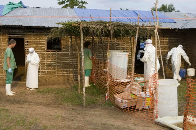
Recommended measures for people caring for those infected with Ebola include the wearing of protective clothing including masks, gloves, gowns, and goggles.[69] The CDC recommends that no skin be exposed.[70] These same measures are recommended for those who may handle objects contaminated by the infected person's body fluids.[71] The CDC in 2014 began recommending that all people receive education on the proper suit-up and removal of personal protective equipment and that someone should be watching each step of the procedure to make sure it is done correctly.[70] In Sierra Leone, the typical training period for the use of such safety equipment lasts approximately 12 days.[72]
The infected person should be in barrier-isolation from other people.[69] All equipment, medical waste, patient waste, and surfaces that may have come into contact with body fluids require disinfection.[71] During the 2014 outbreak kits were put together to help familes treat Ebola in their homes which includes protective clothing as well as chlorine powder and other cleaning supplies.[73] Education of those who provide care in these techniques, and the provision of such barrier-separation supplies has been a priority of the Doctors Without Borders organization.[74]
Ebolaviruses can be eliminated with heat (heating for 30 to 60 minutes at 60 °C or boiling for 5 minutes). To disinfect surfaces, some lipid solvents such as some alcohol-based products, detergents, sodium hypochlorite (bleach) or calcium hypochlorite (bleaching powder), and other suitable disinfectants at appropriate concentrations can be used.[75][76]
Education of the general public of the risk factors for Ebola infection and of the protective measures individuals can take is recommended by the World Health Organization.[1] These measures include avoiding direct contact with infected people and regular hand washing using soap and water.[77]
Bushmeat, an important source of protein in the diet of some Africans, should be handled with appropriate protective clothing and thoroughly cooked before consumption.[1] Some research suggests that an outbreak in the wild animals used for consumption may result in a corresponding human outbreak. Since 2003, such animal outbreaks have been monitored with the aim of predicting and preventing Ebola outbreaks in humans.[78]
If a person with Ebola dies, direct contact with the body should be avoided.[69] Certain burial rituals, which might have included making any kind of direct contact with a dead body, require reformulation such that they consistently maintain a proper protective barrier between the dead body and the living.[79][80] It is recommended that the bodies of people who have died from Ebola be buried or cremated only with proper care.[81] Social anthropologists may help find alternatives to traditional rules for burials.[82]
Transportation crews are instructed to follow a certain isolation procedure should anyone exhibit symptoms resembling the Ebola virus disease.[83] The World Health Organization as of Aug 14, 2014 does not consider travel bans to be useful in decreasing spread.[26]
In laboratories where diagnostic testing is carried out, biosafety level 4-equivalent containment is required, since ebolaviruses are World Health Organization Risk Group 4 pathogens. Laboratory researchers must be properly trained in BSL-4 practices and wear proper personal protective equipment.
Quarantine
Quarantine, also known as enforced isolation, is usually effective in decreasing spread.[84][85] Governments often quarantine areas where the disease is occurring or individuals who may transmit the disease outside of an initial area.[86] In the United States, the law allows quarantine of those infected with ebolaviruses.[87] During the 2014 outbreak, Liberia closed schools.[88] On October 16, 2014, some schools were closed in Ohio and Texas as a precaution after one of two nurses who contracted Ebola (after caring for a person with Ebola) returned to the Cleveland area and may have been on the same plane as some students, teachers, and parents of students from those schools.[89]
Contact tracing
Contact tracing is regarded as important to contain an outbreak. It involves finding everyone who had close contact with infected individuals and watching for signs of illness for 21 days. If any of these contacts comes down with the disease, they should be isolated, tested, and treated. Then repeat the process by tracing the contacts' contacts.[90][91]
Treatment
Standard support
A hospital isolation ward in Gulu, Uganda, during the October 2000 outbreak
No specific treatment is currently approved.[92] However, survival is improved by early supportive care with rehydration and symptomatic treatment.[1] Treatment is primarily supportive in nature.[93] These measures may include management of pain, nausea, fever and anxiety, as well as rehydration via the oral or by intravenous route.[93] Blood products such as packed red blood cells, platelets or fresh frozen plasma may also be used.[93] Other regulators of coagulation have also been tried including heparin in an effort to prevent disseminated intravascular coagulation and clotting factors to decrease bleeding.[93] Antimalarial medications and antibiotics are often used before the diagnosis is confirmed,[93] though there is no evidence to suggest such treatment is in any way helpful. Interferon therapies have been tried as a form of treatment for EVD, but were found to be ineffective.[52]
Intensive care
Intensive care is often used in the developed world.[17] This may include maintaining blood volume and electrolytes (salts) balance as well as treating any bacterial infections that may develop.[17] Dialysis may be needed for kidney failure while extracorporeal membrane oxygenation may be used for lung dysfunction.[17]
Alternative medicine
The Food and Drug Administration (FDA) advises people to be careful of advertisements making unverified or fraudulent claims of benefits supposedly gained from various anti-Ebola products.[94] The FDA has already sent out at least one letter of warning to a seller of colloidal silver who made unverified claims of Ebola related benefits, supposedly derived from the use of their products.[95]
Prognosis
Ebola virus disease has a high risk of death in those infected which varies between 25 percent and 90 percent of those infected.[1][96] As of September 2014, the average risk of death among those infected is 50%.[1] The risk of death was 90% in the 2002–2003 Republic of the Congo outbreak.[97] There are indications based on variations between countries that early and effective treatment of symptoms (e.g., supportive care to prevent dehydration) may reduce the risk of death.[98]
If an infected person survives, recovery may be quick and complete. Prolonged cases are often complicated by the occurrence of long-term problems, such as inflammation of the testicles, joint pains, muscle pains, skin peeling, or hair loss. Eye symptoms, such as light sensitivity, excess tearing, iritis, iridocyclitis, choroiditis, and blindness have also been described.


0 Comments